
NeroVet AI Dental: The Future of Human Oral Care
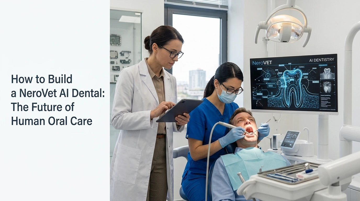
Four seconds. That’s about how long it takes to get a bitewing X-ray to your dentist’s chairside monitor, and then back with 2 highlighted spots; one an area of filling margin and the other a hairline shadow that your dentist may have spotted on a slow Tuesday but would likely slip by on a busy Friday afternoon. Before you’ve rinsed you see the same picture, with plain-English notes.
Quick Facts
- Key Technology: Computer vision, image segmentation and predictive analytics in real time.
- Primary Inputs: Dentists’ X-rays, 3D Cone Beam Computed Tomography (CBCT) scans and intraoral images.
- Key Outputs: Rapid anomaly detection, automated pathology reporting and evidence-based treatment plans.
This is the type of day-to-day maintenance that’s taking place in an increasingly large percentage of dental practices across the nation. There’s no robot arm. No flashy interface. It’s a layer of AI sandwiched between the imaging hardware and the dentist’s eyes, and it takes over the tedious, attention-exhausting, and less efficient tasks that dentists are bad at towards the end of the day.
NeroVet AI Dental is part of this change. The diagnostic engine was originally developed to diagnose dogs and cats with veterinary imaging, where dental disease is common at a very young age and most of the time it is not diagnosed until it is so advanced. It’s the same engine, trained on human radiographic data, and clinically validated, which is now appearing in human practices. And honestly? The crossover is better than one would expect.
How AI is Redefining Human Care
[Digital Scans/X-rays] ➔ [NeroVet AI Analysis] ➔ [Automated 3D Models & Treatment Plan]The core systems pioneered by platforms like NeroVet map directly onto the major shifts happening in human smart dental clinics:
What “AI in Your Dental Chair”
Strip away the marketing and you’re left with three things AI is doing in a working practice today.
- Reading radiographs as they’re captured and flagging suspect regions before the dentist sits down.
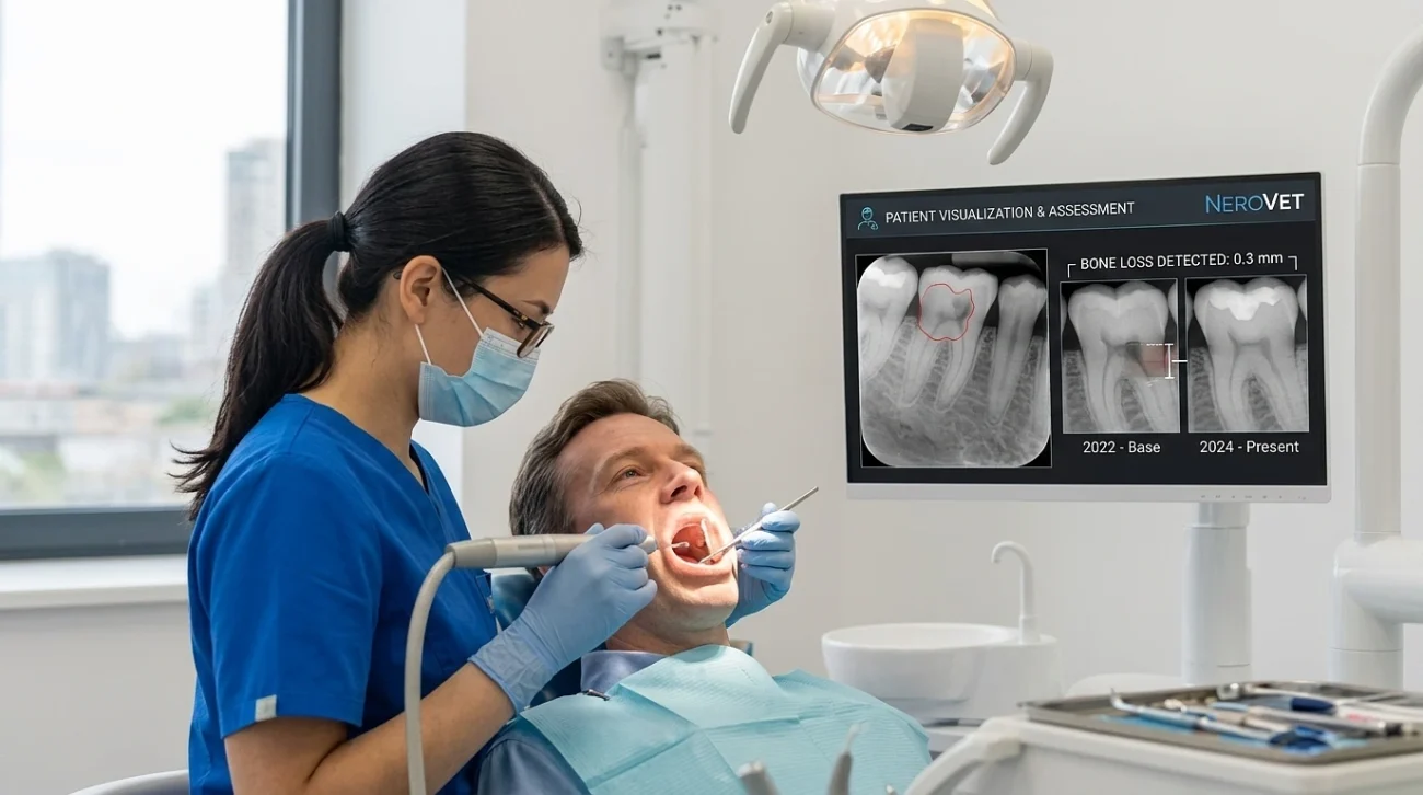
- Tracking pixel-level changes across visits bone loss of 0.3 mm over two years is the kind of drift human eyes lose against memory.
- Turning findings into patient-facing visuals that explain why a procedure is being recommended.
The first one gets most of the press. The other two probably matter more.
Instead of “you have a cavity,” it’s now “this surface has been demineralizing for eighteen months here’s the rate. That’s another warning! Visual explanation, on the other hand, eliminates the trust gap between the dentist’s recommendation and patient acceptance. One of the most persistent challenges in dentistry is treatment acceptance and a clear annotated photograph will get patients past 10 minutes of oral lecture any day of the week.
The only quiet benefit to NeroVet’s engine, it’s history is that it was learned from the crazy arrangement of animal mouths, with breeds and the positioning and shape of teeth all over the place. Edge cases can cause models to fail when constructed solely from the tidy, standardized human data. These are dealt with better by models based on biological chaos.
The clinical Numbers, Without the Hype
Most articles on dental AI fall apart here. They quote “95% accuracy” without saying accuracy at what, on which images, against which reference standard.
Here’s the honest picture from peer-reviewed work in 2024–2025:
In an umbrella meta-analysis of 14 systematic reviews and 20 studies based on the pooled data, AI showed an area under the summary ROC curve of 0.86, sensitivity of 0.85 and specificity of 0.90 for caries detection in 2025. Favorable numbers, but with the note that the data were suggestive of a “controlled study situation”.
The same study, but limited to in-clinic settings, has a different look. A pooled analysis indicated a clinical sensitivity of only 0.35 and specificity of 0.78 for occlusal carious lesions, and a sensitivity of 0.24 and specificity of 0.97 for proximal carious lesions. That discrepancy between research and real world is the same discrepancy that honest vendors should be discussing!
A 2025 panoramic-radiograph review revealed that the best deep learning model achieved a specificity of 0.95, an accuracy of 0.98, and an F1 score of 0.92, with other named, commercial models performing moderate at best.
In periodontal disease, the situation is more likely to be cut and dried. The FDA cleared the Videa Perio Assist, a cloud-based AI system, which automatically measures bone levels in radiographs, turning bone-level analysis with AI into an approved medical device and not a research curiosity in the United States.
The lesson to be learned is not that "AI is perfect" nor is it "AI is overhyped. That is, performance is very task-specific. AI can reliably identify cavitated lesions when bitewings are clear. Not as effective on early non-cavated lesions, on poor quality images and in situations where experts are not in agreement regarding actual positive.The real value of a system like NeroVet is in its consistency. The hour 10 dentist who is a junior will overlook material that a fresh senior will pick up. In published work, AI models, especially the YOLO models and architectures such as DenseNet201 and MobileNetV3, have proven to be high diagnostic accuracy in the detection of cavitated lesions, and some models have been found to perform better than junior dentists. AI doesn’t get tired. That’s a worthwhile task on a hectic agenda.
Why Dental X-rays Are Quietly Becoming Whole Body Screening Tools
This is the part of the AI-in-dentistry story that’s getting too little attention, and it’s the part that matters most for human oral care specifically.
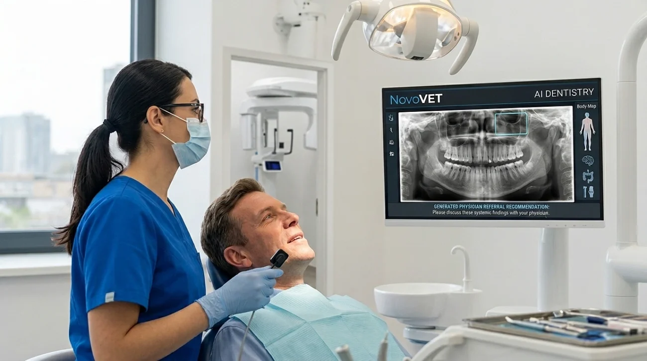
The United States has about 320 million dental radiographs taken each year, and these images typically show evidence of low-grade chronic inflammation and systemic disease such as calcification in the carotid arteries, which is related to stroke. The panoramic images aren’t merely tooth and jawbone. Sinus disease, calcified lymph nodes and bone density patterns that have been linked to osteoporosis are some of the incidental findings they obtain. Until recently, very little of that was recorded in a patient’s medical chart. A dentist is no expert in assessing for stroke risk and, even if they notice something is amiss, the dental-to-medical referral pathway is poorly developed.
AI revolutionizes the economy. The model trained to identify a carotid calcification will be run on all panoramic images at a near zero marginal cost. A recent systematic review has validated that AI can attain high sensitivity and specificity for these calcifications on conventional panoramic radiographs, but the “traditional workflow” is not often followed by actionable referrals. The dentist is not required to be a cardiologist. All they have to do is to get a generated note to make the sentence “please discuss with your physician” appear in the report.
The clinical case is good. The American Heart Association scientific statement on periodontal disease and atherosclerotic cardiovascular disease highlighted the need for regular dental screenings and targeted periodontal treatment to help reduce systemic inflammation, especially in individuals at high risk for atherosclerotic cardiovascular disease (ASCVD) who require it. Affecting 20–50% of the adult population worldwide, periodontal disease is a chronic inflammatory condition which is linked to endothelial dysfunction, which is also involved in heart attacks and strokes.The mouth has long been considered an indicator of systemic health. AI is the first technology that makes it affordable and feasible to see through that window on every patient, every visit not just when they’ve already got something that’s gone wrong.
What the Technology Still Gets Wrong
Any honest piece on dental AI has to name the failure modes.
- The most common complaints of practicing dentists are false positives. A model that’s tuned for sensitivity is going to identify something that doesn’t really have a lesion, and that’s a stain that looks like a lesion, or a shadow that looks like bone loss, but isn’t. Eventually this leads to a loss of trust by the clinician (“the AI cries wolf”) or to overtreatment by one with less experience who submits to the algorithm too easily.
- The problem is deeper, with respect to dataset bias. The vast majority of the dental AI models published were built from imaging data from limited populations, typically university clinics from only a few countries. Restorations may perform down, often significantly, on patients whose dental anatomy, restoration pattern or imaging hardware differs. If a model is successful at 92% accuracy in Helsinki, it may be only 78% accurate in Houston and you wouldn’t know unless somebody observed.
There’s also the vendor demo lack of integration issue that nobody speaks of. Dentrix, Eaglesoft, Open Dental and various other legacy software are the norm in dental practices, and adding an AI overlay to communicate clearly with existing radiographic equipment, EHRs, and billing systems is no easy task. Any practice which purchases an AI solution and fails to designate someone to actually manage the integration finds it languishing in a year.
NeroVet AI Dental, like every other player in this space, has to be judged on how it handles these specific things.